
REDMOD: AI spots pancreatic cancer on CT scans more than a year early
Pancreatic cancer AI model REDMOD found warning signs on CT scans 475 days before diagnosis on average, a study published in the journal Gut found.
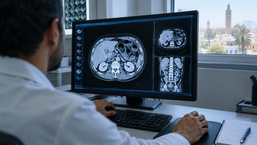
Illustrative image
Routine CT images that radiologists had judged normal already carried traces of pancreatic cancer, and an artificial intelligence model called REDMOD was able to read them. A study published online in the journal Gut found the tool picked up these signals 475 days, on average, ahead of a clinical diagnosis.
Its sensitivity reached 73%. Radiologists with years of experience, reading the same images, identified only 39% of the patients who would go on to develop the disease, meaning the software caught close to double their share.
Reading tissue texture
The name stands for Radiomics-based Early Detection MODel. It was built to find the most frequent form of the disease, pancreatic ductal adenocarcinoma, by analysing radiomics: minute texture patterns in the organ that the human eye struggles to see on standard computed tomography.
The software also finds and outlines the pancreas automatically, isolating it from neighbouring organs and tissue. Because no specialist has to draw the boundaries manually, differences between one reader and another are avoided.
Survival from this cancer is low, chiefly because it tends to be caught late. In its early phase it produces few symptoms, the changes it causes are often too slight for ordinary imaging, and it can spread fast.
219 patients, 1,243 controls
The researchers gathered 219 patients from several hospitals whose abdominal scans had shown no sign of cancer to radiologists. Every one of them was diagnosed with pancreatic cancer later on.
The gap between scan and diagnosis varied. It was 3 to 12 months for 87 people (40%) and 12 to 24 months for 76 (35%). For the remaining 56 (25%), imaging took place over 24 months beforehand, in some cases close to three years. Tumours formed in the pancreatic head in 64% of patients.
This group, with an average age of 69 and ages from 34 to 88, was set against 1,243 people matched by age, sex and scan date. None of them developed the disease during as long as three years of follow-up. Their mean age was 64, within the same 34-to-88 range.
“This temporal window holds profound significance, as attaining such early detection would substantially augment the probability of cure and improved survival,” the researchers wrote.
Citing modelling work, they said that lifting the proportion of tumours found while still localised, from 10% to 50%, would leave survival rates more than twice as high, and argued that timing of diagnosis is the biggest single factor in outcomes.
Wider gap on older scans
REDMOD’s lead grew when the images predated diagnosis by over two years. On those, the model found 68% of cases, almost triple the radiologists’ 23%.
The team also checked how it handled people without cancer. Across an independent set of 539 patients drawn from several hospitals, slightly more than 81% were correctly classed as cancer-free. On NIH-PCT, a public collection of 80 patients from the US National Institutes of Health, the rate rose to 87.5%.
Repeat scans of the same individuals, taken several months apart, produced the same verdict 90% to 92% of the time. The authors took this stability as further support that the model was tracking genuine disease-related changes, years before a diagnosis in some patients.
Further testing needed
The researchers say more work is required before routine clinical use. Ethnic diversity in the study population was limited, which could influence results in other groups.
They also call for prospective trials, among people at higher risk, such as those with unexplained weight loss or diabetes diagnosed recently, to establish whether the tool works in everyday practice.